
Dr Klaus Schmitt
Well-known member
A while ago I had the honor to work with Neal Larson (the guy who found SUE the famous T-Rex) on a paper on fossilized cephalopods found in Hajoula, Lebanon. LINK to Paper
Today it is about enhancing the visibility of fossil bone and tissue structures using UV reflected and UV stimulated visible fluorescence photography. I will also use my remapping technology consisting of a visible image, a reflected UV image and an UV stimulated visible fluorescence image for that and combine them into multispectral images.
Lens used was my CERCO quatz fluorite lens, light sources were a modified high power Xenon flash as well as a NICHIA 365nm Power LED. Target was a fossilized fish from Solnhofen, Germany, approx. 100 Mio years old.
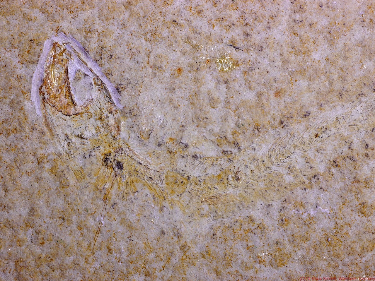
Visible light image using UV/IR Cut filter:
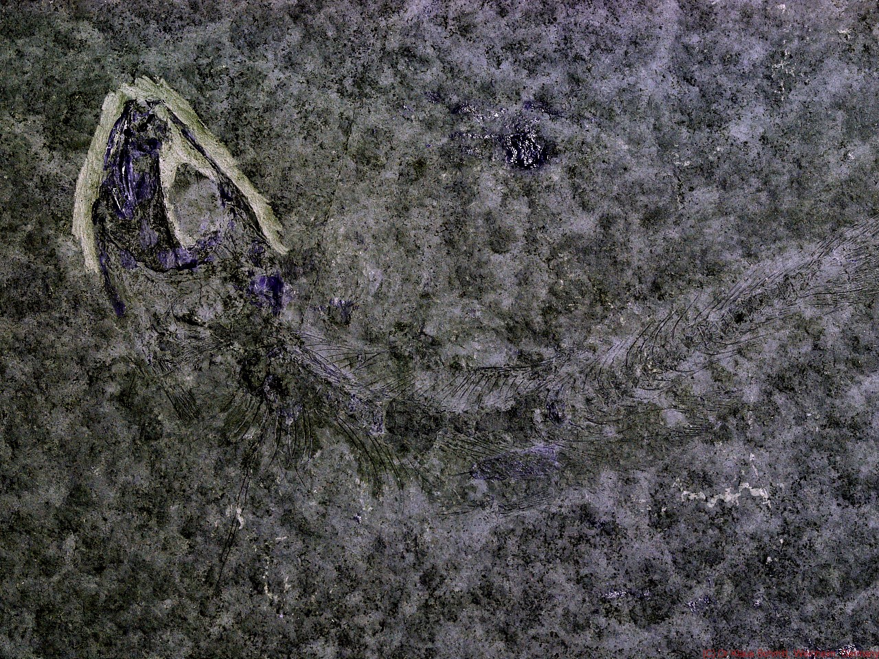
Reflected UV image using Baader-U filter (310-390nm):
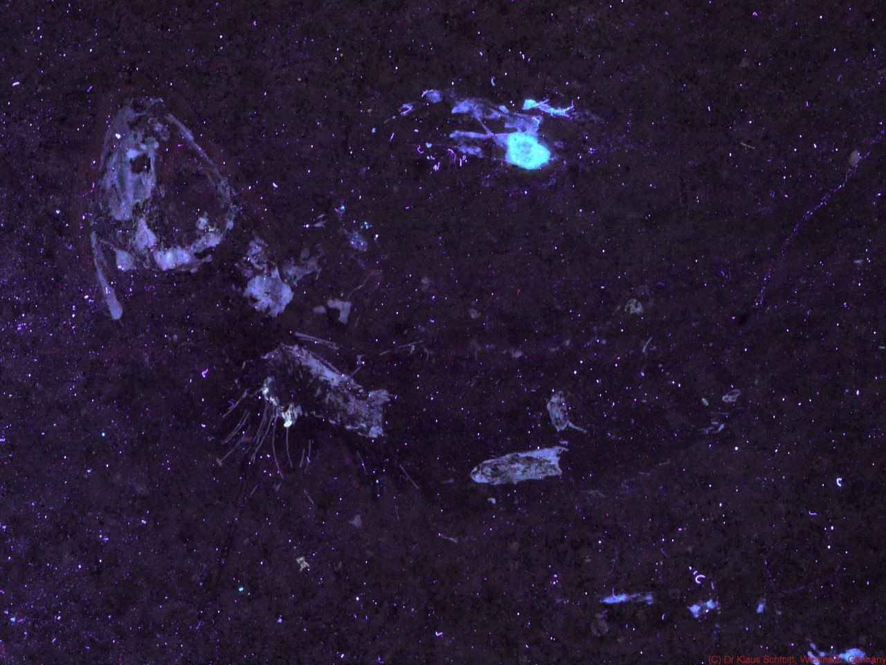
UV stimulated visible fluorescence (FL) using Nichia 365nm UV LED:
Combined VIS - FL multispectral image:
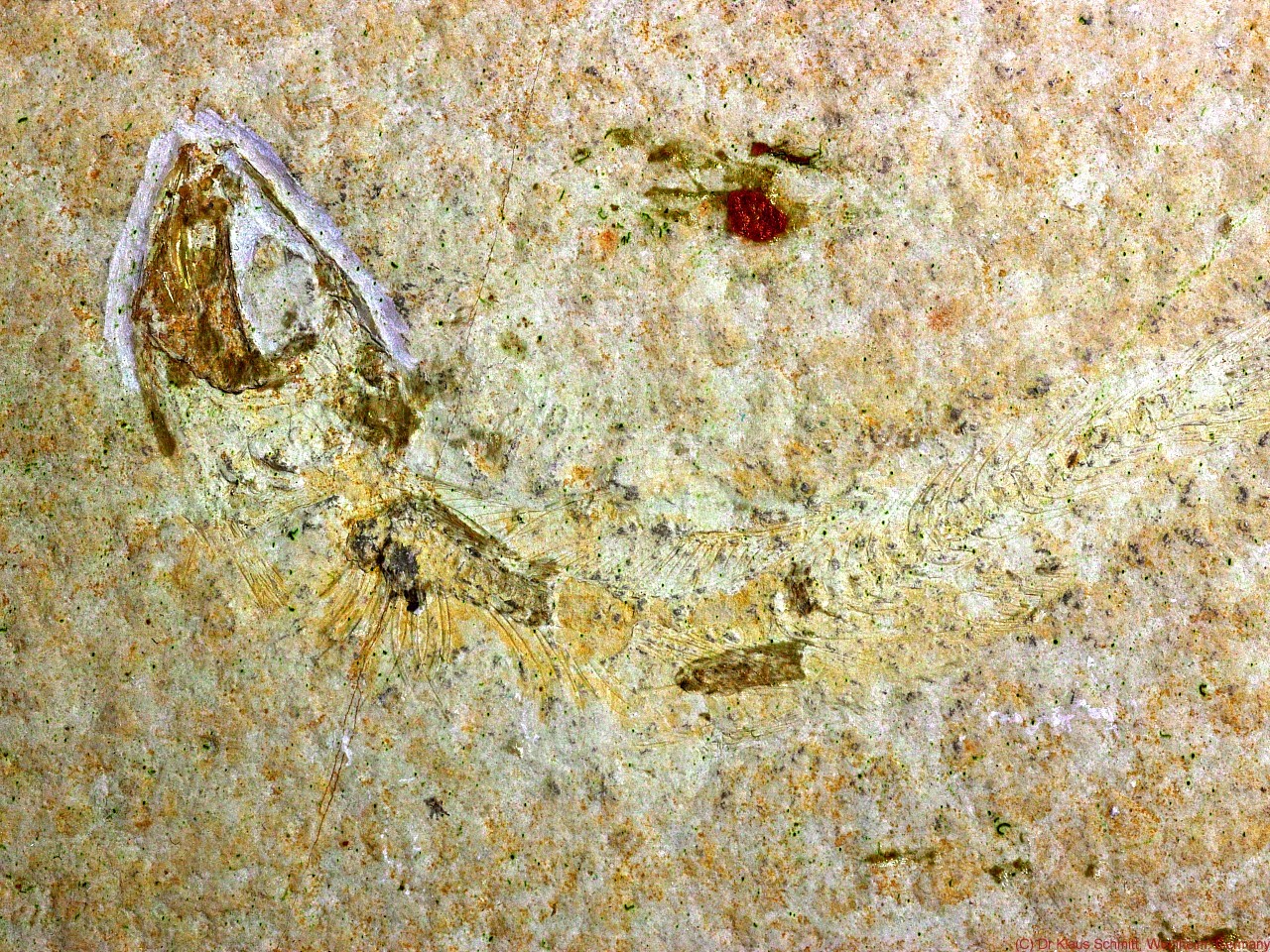
Combined VIS - UV multispectral image:
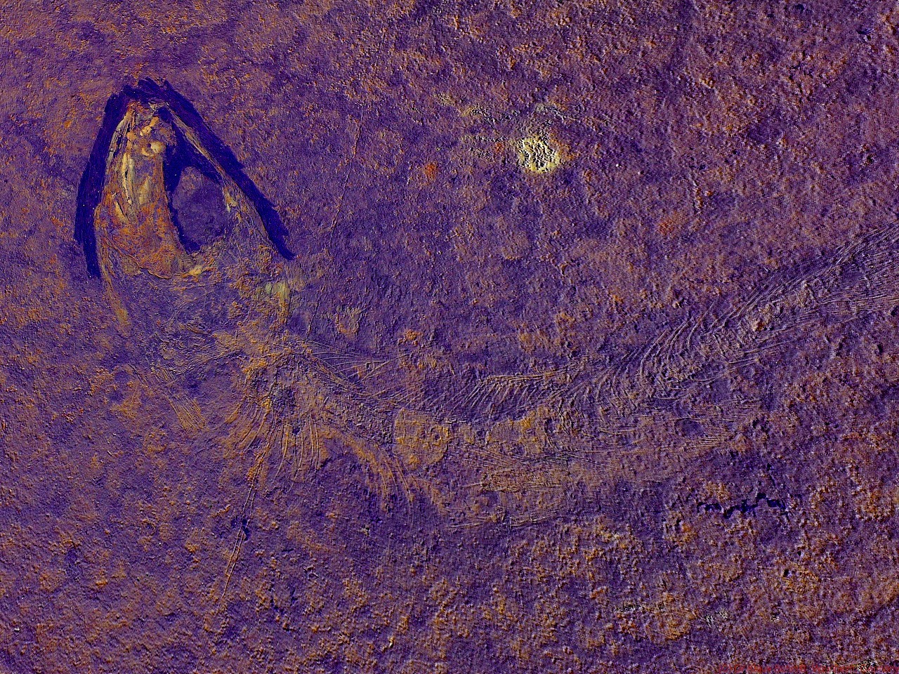
Combined FL - UV multispectral image:
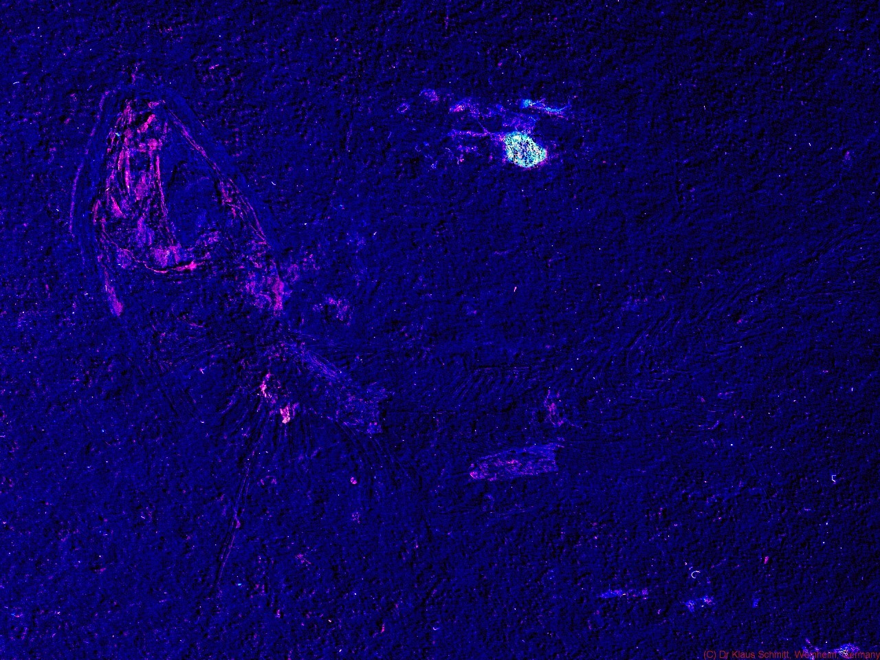
It gets nicely visible that using UV light brings out much more details than normal visible light photography and by doing so enhances the visibility of preserved bone and tissue structures quite a bit. Combining those different images even more enhances the structures.
So Long, and Thanks for All the Fish....
Today it is about enhancing the visibility of fossil bone and tissue structures using UV reflected and UV stimulated visible fluorescence photography. I will also use my remapping technology consisting of a visible image, a reflected UV image and an UV stimulated visible fluorescence image for that and combine them into multispectral images.
Lens used was my CERCO quatz fluorite lens, light sources were a modified high power Xenon flash as well as a NICHIA 365nm Power LED. Target was a fossilized fish from Solnhofen, Germany, approx. 100 Mio years old.
Visible light image using UV/IR Cut filter:
Reflected UV image using Baader-U filter (310-390nm):
UV stimulated visible fluorescence (FL) using Nichia 365nm UV LED:
Combined VIS - FL multispectral image:
Combined VIS - UV multispectral image:
Combined FL - UV multispectral image:
It gets nicely visible that using UV light brings out much more details than normal visible light photography and by doing so enhances the visibility of preserved bone and tissue structures quite a bit. Combining those different images even more enhances the structures.
So Long, and Thanks for All the Fish....
Last edited: